|
|
Information box |
The main purpose of this site is to extend the
intraoperative monitoring to include the neurophysiologic
parameters with intraoperative navigation guided with Skyra 3
tesla MRI and other radiologic facilities to merge the
morphologic and histochemical data in concordance with the
functional data.
 CNS Clinic
Located in Jordan Amman near Al-Shmaisani hospital, where all
ambulatory activity is going on.
Contact: Tel: +96265677695, +96265677694.
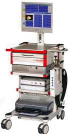
Skyra running
A magnetom Skyra 3 tesla MRI with all clinical applications
started to run in our hospital in 28-October-2013.
Shmaisani hospital
The hospital where the project is located and running diagnostic
and surgical activity. |
|
 |
|
|
 |
SYNGO.VIA |
 |
 syngo.via is the new imaging software,
creating an exciting experience in efficiency and ease of use –
anywhere. syngo.via is your agent for productivity throughout
your radiology workflow. No other solution supports and
integrates all MR tasks in a comparable way – from planning and
scanning to result sharing.
syngo.via is the new imaging software,
creating an exciting experience in efficiency and ease of use –
anywhere. syngo.via is your agent for productivity throughout
your radiology workflow. No other solution supports and
integrates all MR tasks in a comparable way – from planning and
scanning to result sharing.
Integrated Engine concept
The Engine concept integrates the scanning and reading processes
into one holistic workflow and enables you to maximize your
scanner and application investment.
Key features The Dot (Day optimizing throughput) Engines
optimize the performance of MAGNETOM Skyra and offer patient
personalization, user guidance, and exam automation.
syngo.via offers reading workflows, which are optimally adjusted
to the Dot Engines for the best reading outcomes
The scanning and reading workflows are easily customizable to
the user’s standards of care.
Results from the scanner are optimally displayed in the
reading workflows.
Key benefits Guidance, standardization and flexibility is
offered for every step of the workflow, reducing the need for
further inquiry
Increasing throughput, minimizing recalls, and enhancing quality
of care.
Networked MR scanner – the scanner workplace for optimized
productivity
syngo.via handles images from any MR system. With MAGNETOM
scanners, the integration is perfect. The networking
capabilities of the MAGNETOM scanners and syngo.via will
transform the technologist workplace of today. One consistent
workflow is offered, from planning, to scanning to viewing, and
processing.
Key features Host computer of the MAGNETOM Skyra is
rich-thin-client enabled. The syngo.via client can be run with
only one keyboard and one mouse – no cumbersome change of
devices necessary.
syngo.via offers unique workflows to support the technologist:
• Check Protocol: pre-defined protocols are automatically
displayed
• Initiate Scan: get guidance by additional text info for the
optimal scan and patient set-up. Start patient registration
directly out of syngo.via
• Check images: immediate availability of images, easy quality
check of images
Automatic transfer of the patient data and planned protocols
from syngo.via to the scanner. No need for double entry of
information.
Automatic selection of the appropriate syngo.via reading
workflow
syngo.via and MAGNETOM Skyra UI – both based on the proven syngo
UI concept.
Key benefits
Work with different patients side-by-side without any screen
overlays and possible confusions. E.g. begin with patient
registration of one patient, while other patient is still
scanned.
Automatic transfer of information – reduced need for
clarifications
Higher throughput, reliable results, and reduced costs.
Pre-requisites Additional monitor and one syngo.via license
Direct Protocol Transfer (DPT)
syngo.via redefines protocol management – providing remote
protocol planning, and automated selection
of the right protocols at the scanner. syngo.via offers a
dedicated workflow for protocol planning and
distribution. The radiologist can plan the protocols from
anywhere in the institutional network.
Key features Remote protocol planning from any syngo.via client
to select or modify a planned
protocol (and perhaps add further explanations) for a patient
examination before
the patient is registered at the MR system. The planed protocol
will be automatically
transferred to the scanner via DICOM Modality Worklist.
The technologist works with the same syngo.x view directly at
the scanner –
accessing the same information, without any further handwritten
notes or need for
clarifications.
Automatic transfer of the patient data and planned protocols
from syngo.via to the
scanner. No need for double entry of information.
In addition: Remote Protocol distribution:
• upload, change and/or delete any protocol from your MAGNETOM
scanners
(available for all Tim Systems)
• send examination protocols to every other connected scanner
Key benefits Define standards of care and easily distribute the
related protocols among your Tim
systems.
Save time for clarifications – while minimizing re-scans
Easy, automated, and efficient protocol handling – from anywhere
DPT works with MAGNETOM Skyra and any other Tim system.
Direct Image Transfer (DIT)
After completion of a series of images, they are transferred
automatically to syngo.via. Easily view the
images with any syngo.via client immediately after they were
acquired.
Key features This data will be automatically transferred to the
syngo.via data base and loaded
into the related workflow. Enhanced DICOM MR enables to transfer
fMRI data and
spectroscopy raw data in new DICOM standard format. In syngo.via
and any PACS
supporting this standard, these data can be handled.
Key benefits Images are immediately available throughout the
institution. This enables fast and
convenient feedback from everywhere.
Direct Image
Transfer Pro
A direct cable connection is build between the MAGNETOM scanner
and the
syngo.via server. This ensures a guarantied performance for
image transfer.
Seamless workstation integration
syngo.via. integrates smoothly with syngo MultiModality
Workstation (MMWP). Open MMWP directly out of syngo.via, and
vice versa.
Key features Remotely open the same patient at the MMWP easily
with syngo Expert-i
The MMWP results can then be easily integrated into the
syngo.via report
syngo.via client can be opened from any MMWP with one click
Key benefits Remote and easy access of all MMWP applications
Smooth integration of results into syngo.via
 |
syngo. MR General
Engine |
 |
A rich suite of functionality to cover all of your routine
reading and post processing needs. Whatever you need to read,
find the right tools, right at hand.
Features
•MR Radiology Workflows: predefined layouts for Head, C-Spine,
T-Spine, L-Spine, Whole Spine, Breast, Prostate, Abdomen, Hip
and Knee scans.
•MR Cardio-Vascular Workflows: Single Station Angio, Multi
Station Angio, Angio TimCT, Angio TWIST, Cardiac Reading
•Subtraction, MeanCurve
•Workflow optimized report template included.
•Disease specific report templates for breast (according to
BIRADS Report) and prostate
•MR Misc data: collection of additional data, that have not been
loaded in any of the predefined layouts (e.g. additional scans,
normally not part of the workflow). A single click that makes
sure no data were missed by the user.
Clinical Applications
standard MRA, MR Mammography, Prostate, Neuro and Ortho Reading
 |
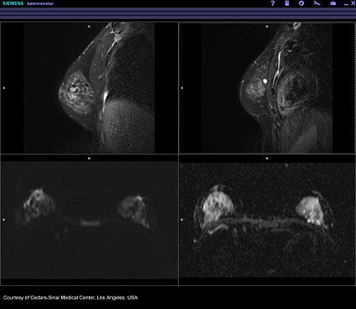 |
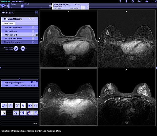
| Dual monitor. Left: Breast Reading |
Dual monitor. right |
 |
 |
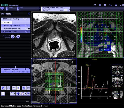
| Dual monitor. Left: Prostate Reading |
Dual monitor. right |
Additional Information
My cases, ready. Whether it is the 3D Reference Point, the Auto
zooming functionality in multi stage exams, or mean curve and
subtraction, the syngo.MR General Engine extends syngo.via by
adding software for advanced and routine MR radiology usage. It
includes workflows for dedicated MR examinations that load and
structure examination results automatically into meaningful
layouts, including user support to make sure that no data is
missed.
The perfect match to such applications as the Brain Dot Engine;
Knee Dot Engine; Abdomen Dot Engine, TWIST, TimCT Angio, and
Inline Composing, the syngo.MR General Engine enables an
optimized workflow from scanning, to processing, to reading.
|
|
Networked Scanner |
|
Modalities and IT become one, within the institution and beyond.
Features
Our Networked Scanner offers you flexibility:
At the individual case level use syngo.via* to change protocols
for a specific case.
At the enterprise level standardize and efficiently manage
protocols across your MAGNETOM systems.
With Networked Scanner, the radiologist / lead technologist can
adjust the scans assigned from the modality work list, and
ensure that the patient receives the scan she needs.
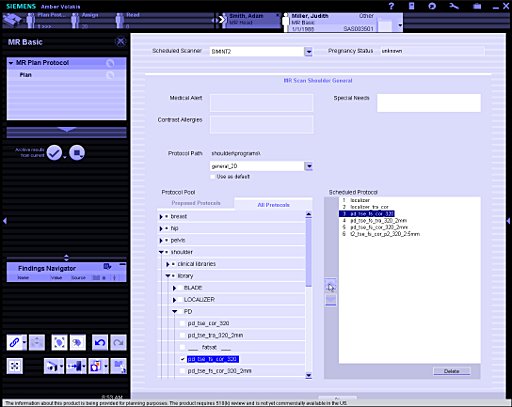 |
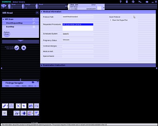 |
| Patient Protocol Planning |
Patient Protocol at the scanner |
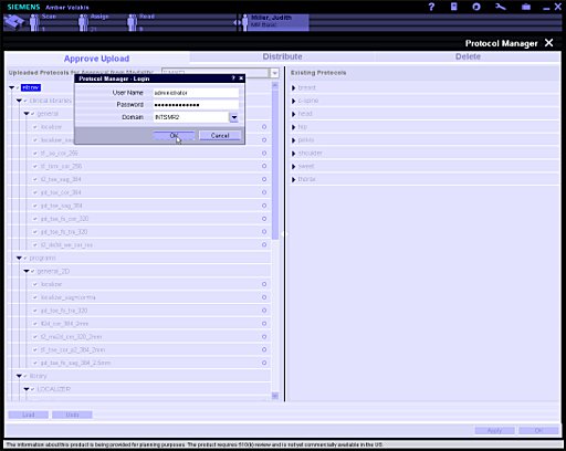 |
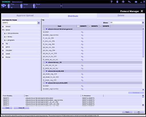 |
| Protocol Management Upload |
Protocol Management Distribution |
The operator also at any time can optimize the images and
adjust strategies during the scan and then immediately
manipulate and read the images in syngo.via. This ensures that
the radiologists receive the images they need to complete the
diagnosis.
Share protocols across the enterprise.
Have a complete overview of all protocols on all systems from
any workplace.
Reduce scan downtime when managing protocols.
Match the patient to the right scanner.
Networked scanner enables all of these activities and helps to
ensure that the organization receives the structure that it
needs.
|
|
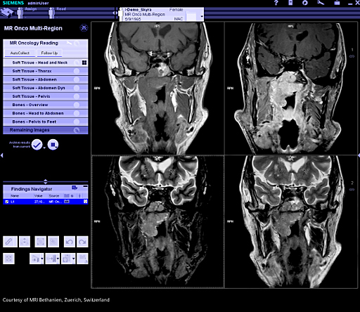
syngo.MR Onco Engine |
|
Make oncology diagnosis fast, intuitive and more robust.
Features
syngo.MR Onco Engine combines features that allow for efficient
oncological reading and reporting.
Included Workflows: Onco Multi-Region, Onco Brain, Onco Liver,
Onco TimCT
syngo.MR 3D Lesion Segmentation provides convenient volumetric
evaluation of lesions.
Onco Reporting: Each workflow contains a dedicated reporting
template and oncological findings classification - including a
pictogram for visual information transfer to the referring
physician to quickly localize spots of interest. Total tumor
load can be calculated. Response calculations comply with RECIST
and WHO Response Criteria.
 |
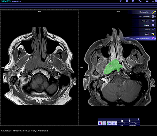 |
| Dual monitor, left: Soft Tissue |
Dual monitor, right: 3D Lesion
Segmentation |
 |
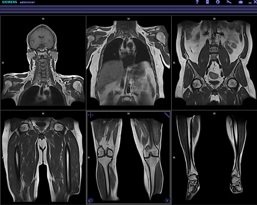 |
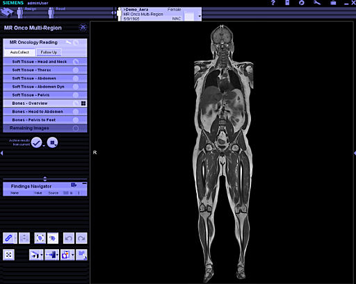
| Dual monitor, left: Whole-body
Overview |
Dual monitor, right: Whole-body
Overview |
Additional Information
syngo.MR Onco workflows structure large amounts of data
automatically and quickly into layouts focused on oncology
reading. Each workflow contains a dedicated follow-up reading
layout (optimized for dual monitor support).
The 3D Lesion Segmentation is particularly useful for oncology
applications (e.g. volumetric evaluation of tumors, lymph nodes
and metastases), but also for non-oncology lesions with
sufficient contrast to surrounding tissue. Intuitive editing
tools allow adjustment to the segmentation if necessary.
The perfect match to TimCT Onco and the syngo.MR Onco Engine
enables an optimized workflow from scanning, to processing, to
reading.
|
|
syngo.MR Neuro
Perfusion Engine |
|
With every tool you need for fast and standardized diagnoses, we
are establishing a whole new level of speed and flexibility in
acute neurology.
Features
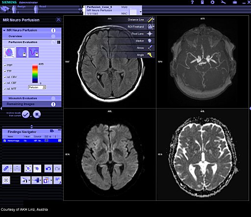
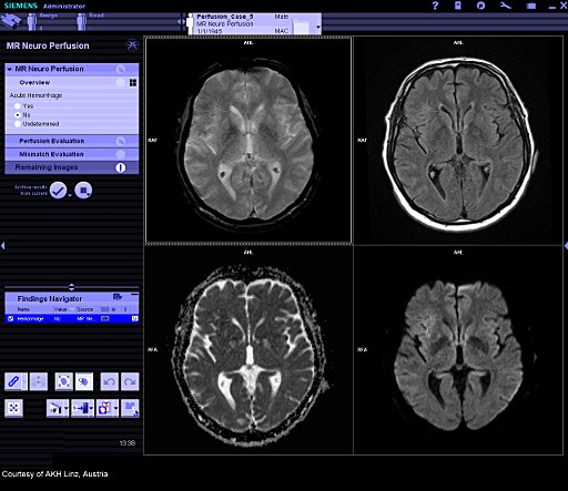
The syngo.MR Neuro Perfusion Engine bundles three Neurology
features for detailed brain assessments: MR Neuro Perfusion
Evaluation, Automatic local AIF calculation and
Perfusion-Diffusion Mismatch calculation.
color display of the relative Mean Transit Time (relMTT)
relative Cerebral Blood Volume (relCBV) relative Cerebral Blood
Flow (relCBF)
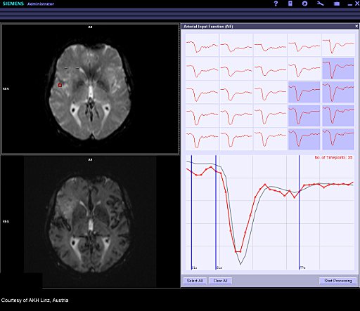
Flexible selection of the Arterial Input Function (AIF) for more
reliable analysis taking into account the dynamics over time of
the contrast agent enrichment.
Additional maps such as TTP, corrCBV
Automatically calculates local Arterial Input Function (AIF) and
generates the perfusion results.
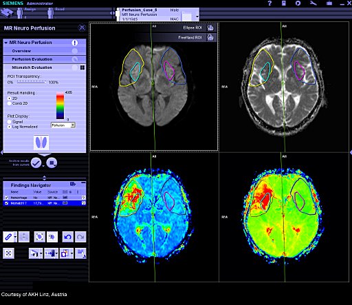
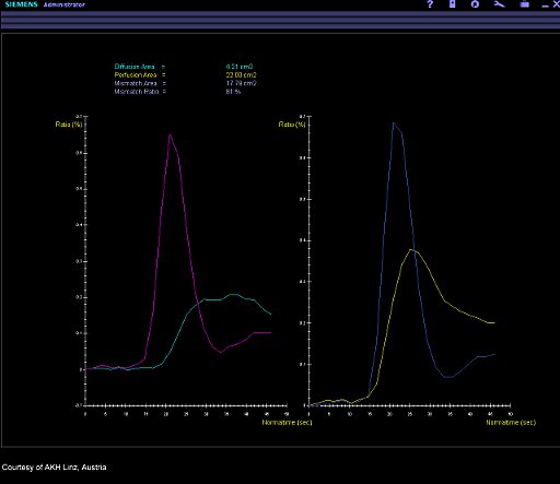
MR Neuro Perfusion Mismatch: calculates the Perfusion-Diffusion
difference.
Disease oriented MR Neurology reporting template included.
 |
 |
| Dual monitor, left: Perfusion
Evaluation |
Dual monitor, right: Perfusion
Evaluation |
 |
 |
| Dual monitor, left: Mismatch
Evaluation |
Dual monitor, right: Mismatch
Evaluation |
 |
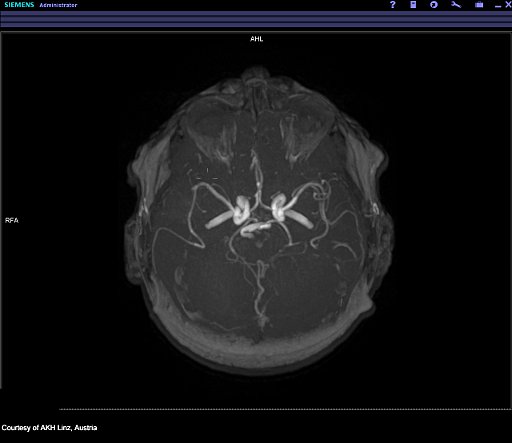 |
| Dual monitor, right: Overview |
Dual monitor, right: Overview |
Additional Information
My cases, ready
Shorten reading preparation: Images are mapped to predefined
reading steps that you can determine. You no longer have to
search for and organize your images. Ensure that your reading
happens on the same set of images every time.
Structured Reading: Standardized Diagnosis. Ensure that your
team reads the same images the same way to ensure consistent
outcomes. Images and diagnostic steps cannot be forgotten as
long as one follows the structured workflow.
Compile your information automatically into a report, whose
template suits your needs.
My places, networked
With its client server technology, reading is possible from
anywhere* in your organization. Time is brain. Doctors should
spend time reading, not walking to a work station.
|
|
syngo.MR Cardiac 4D
Ventricular Function |
|
Our „hands-off“ post processing allows you to see immediate
results when you open your case.
 |
 |
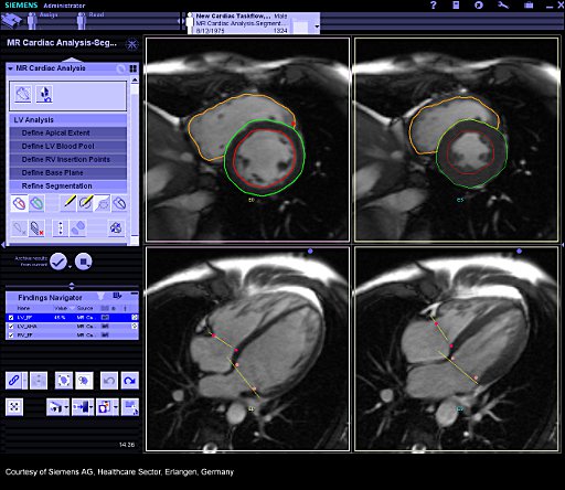
| Dual monitor, left: Segmentation |
Dual monitor, right: Gallery |
|
Features
Instant display of preprocessed segmentation (left ventricle)
based on prize-winning algorithm;
Automatic Calculation of ventricular volumes, ejection fraction
and myocardial mass
Automatic Calculation of myocardial thickening for all
myocardial segments
Semiautomatic processing of the right ventricle
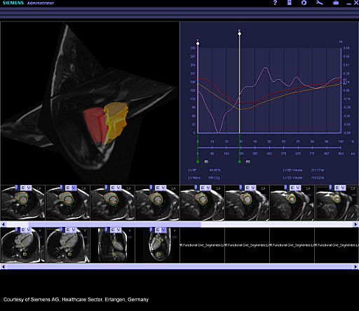
Dynamic evaluation such as time-volume diagrams, filling rate
and myocardial wall motion
Integrated and synchronized movie display
Graphical display of results (tables, bull's-eye plot, 4D model
of the heart)
Integration of the results in a report for documentation
Clinical Applications
Functional and volumetric evaluation are the cornerstone of
every cardiac MR examination in ischaemic heart disease as well
as in cardiomyopathies such as myocarditis. Precise analysis of
all relevant volumetric parameters such as ejection fraction,
stroke volume and e.g. segmental analysis of wall thickening.
Additional Information
My cases, ready. See images presented by type or anatomical
view. syngo.via automatically recognizes and sorts them into
predefined layouts, preventing mix-up of images by order or
location.
Easily review automatically created short and long axis contours
with syngo.via’s* guided approach. The software calculates all
relevant parameters for cardiac functional analysis and displays
them in standardized graphs and tables – from ejection fraction
(EF) to regional wall thickening, among others
The perfect match to the Cardiac Dot Engine and Inline VF,
syngo.MR Cardiac 4D VF enables an optimized workflow from
scanning, to processing, to reading. |
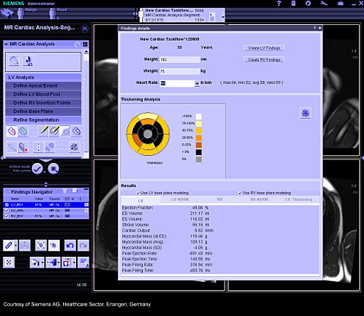 |
| |
Dual monitor, left: Report Finding
details |
|
|
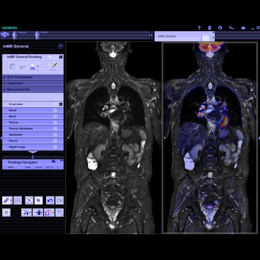
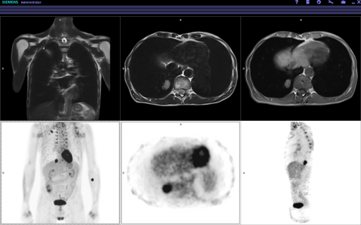
syngo.mMR General
Engine |
|
Biograph mMR and syngo.via* are one. For the first time,
Biograph mMR provides MR and PET data as one dataset - molecular
MR acquisition data. Every Biograph mMR includes syngo.mMR
General to fully utilize this one dataset in your clinical
environment.
Features
Automatic loading of mMR data
Visualization in precise registration
Direct Fusion
Lesion propagation between MR and PET
Support of multiple time-point analysis
Summary of the final result in one report
 |
 |
| left side of dual-monitor display |
right side of dual-monitor display |
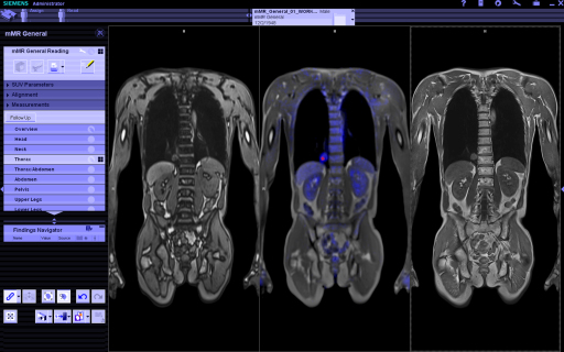 |
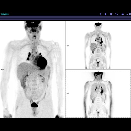
 |
| left side of dual-monitor
display |
right side of
dual-monitor display |
Additional Information
My cases, ready.
Take full advantage of identical Frames of Reference:
Data preparation reduced
MR and PET Markings correlated automatically
Findings linked exactly in the Findings Navigator
Comprehensive set of evaluation tools
Easy creation of findings
My places, networked.
See what you need, where you need it**, how you need it:
Network-wide access for viewing, reading, and collaboration
Collaboration function allows two experts to diagnose the same
case at the same time
With suspend/resume, one doctor is notified when the other has
finished reporting
|
|
|